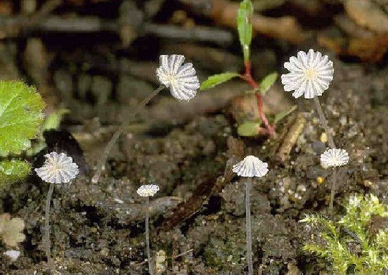
 Closed pileus 4 mm high and 3 mm broad, oval to ellipsoid then conical to convex, finally flat with upturned split margin, pale grey-brown with yellow-orange centre of pileus; with granular-powdery veil; expanded pileus 4-8 mm, slowly wilting; lamellae narrowly adnate, whitish to blackish; stipe 12-18 x 0.4-0.8 mm, hollow, equal, when young pruinose, base with white velar flocks; smell absent. Closed pileus 4 mm high and 3 mm broad, oval to ellipsoid then conical to convex, finally flat with upturned split margin, pale grey-brown with yellow-orange centre of pileus; with granular-powdery veil; expanded pileus 4-8 mm, slowly wilting; lamellae narrowly adnate, whitish to blackish; stipe 12-18 x 0.4-0.8 mm, hollow, equal, when young pruinose, base with white velar flocks; smell absent.
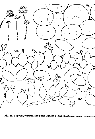
Spores 7.5-11.5 x 6-7.5 x (5-)5.5-6.5 µm, very variable in size (see discussion below), oval or ellipsoid in frontal view, the broader ones sometimes a little angular, ellipsoid to slightly amygdaliform in side view, with small central germ pore, c. 1.2 µm in diameter; basidia 4-spored; cheilocystidia (sub)globose, ellipsoid to ovoid, 10-21 in length or with diverculations and then up to 30 µm in length; pleurocystidia absent; pileipellis made up of slightly thick-walled, ventricose, ellipsoid and subglobose elements, 10-21(-29) µm wide. Clamp-connections not found.
|
Remarks |
The description and illustration given here is based on the original publication of Bender l.c.
Coprinus ramosocystidiatus can be easily identified by the diverticulate cheilocystidia. The only other species known in subsect. Nivei with such cystidia, is C. cordisporus which has completely differently shaped spores. From the type-locality several collections were gathered. Spore-size appears to be very variable: coll. 7 Aug. 1987: (8.5)10-11.5 x 6-7 x (5-)5.5-6 µm, coll. 15 Aug. 1987 (holotype): 7.5-8.5 x 6-7 x 5.5-6 µm, coll. 20 Aug. 1987: 9-10.5 x 6.5-7.5 x 5.5-6.5 µm.
Although Enderle & Bender (l.c.) placed C. ramosocystidiatus in the 'micaceus'-group, we place it in subsect. Nivei, on account of the mealy-powdery aspect of the veil, which can be easily seen in the coloured photograph that was published in the original publication. It is possible that the caulocystidia described by Bender actually represent remnants of the veil, adhering to the stipe surface. In the illustration (Bender, l.c.), these 'caulocystidia' were erroneously named 'pleurocystidia.' |